
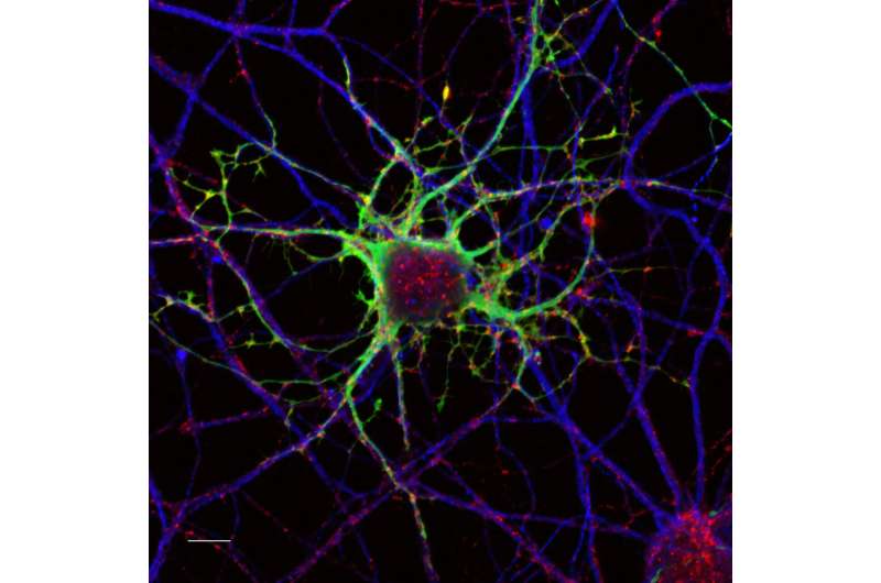
Researchers observed a decrease in nerve cell density in periosteum of HFD-fed mice. Credit: Cactus Communications
Diabetes Mellitus is a chronic metabolic disorder and is one of the leading chronic diseases worldwide. It is widely known for its impact on blood sugar levels and conditions related to the cardiovascular system, kidneys, eyes, and nerves.
One of its most common and crippling complications is diabetic peripheral neuropathy (DPN), which is characterized by loss of nerve fibers, impaired sensation and pain, especially in the limbs. While these effects are commonly known, a lesser-known consequence is its effect on bone health, characterized by decreased bone mineral density with an increased risk of fractures.
Recent research suggests that DPN could be linked with an increased fracture risk. However, the exact biological connection between diabetic nerve damage and skeletal health remains underexplored. Bridging this gap, a team of researchers led by Dr. Aaron James from Johns Hopkins University, Baltimore, U.S., revealed a direct connection between DPN and bone degeneration, linking it to reduced cell signaling. The findings of the investigation were published online in the journal Bone Research on July 4, 2025.
To observe this link, the team modeled type 2 diabetes in young male mice using a high-fat diet (HFD) and observed the classic signs of metabolic dysfunction like weight gain, insulin resistance and elevated blood glucose. In addition to these, the mice also developed a measurable level of nerve damage (neuropathy) indicated by a decrease in nerve fibers in the outer skin layer and reduced response to pain stimuli. Moreover, the researchers also observed a striking loss of nerve fibers in the bones themselves.
Notably, the longer bones of the HFD-fed mice showed up to a 76% reduction in nerve densities. This reduction in nerve density also coincided with weakened bone structure, including a reduction in bone volume, cortical (outer bone) thickness as well as trabecular (inner spongy bone) density.
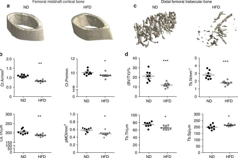
High-fat diet (HFD) feeding results in cortical and trabecular bone alterations. Normal diet (ND) or HFD feeding in C57BL/6J mice was instituted in week 4 of life, with analysis at week 16 of life. a µCT images of femoral midshaft cortical bone. b µCT quantifications of cortical area (Ct.Ar), cortical perimeter (Ct.Pm), cross-sectional thickness (Cs.Th) and polar moment of inertia (pMOI). n = 8 mice per group. c µCT images of distal femoral trabecular bone. d µCT quantifications of Bone volume per total volume (BV/TV), Trabecular thickness (Tb.Th), Trabecular number (Tb.N) and trabecular separation (Tb. Sp). n = 8 mice per group. Graphs represent average values ± 1 SD, *P < 0.05, **P < 0.01 and ***P < 0.001. Comparisons between groups were analyzed by unpaired Student’s t test. Credit: Bone Research (2025). DOI: 10.1038/s41413-025-00436-x
"We've known that patients with diabetes have a higher risk of fractures, but our study shows that part of this risk may come directly from disrupted nerve-bone communication, " comments communicating author, Dr. James.
To uncover the underlying biological mechanism, the team conducted single-cell RNA sequencing and analyzed both sensory neurons as well as periosteal cells—thin layers of cells that surround the bones and are critical for growth and repair. They observed that a group of signaling molecules like VEGFA (Vascular Endothelial Growth Factor A), BDNF (Brain-Derived Neurotrophic Factor) and CGRP (Calcitonin Gene-Related Peptide) secreted by healthy neurons, interact with periosteal cells to promote bone formation and repair.
But under diabetic conditions, this nerve-to-bone signaling was impaired, and instead of forming new bone, the periosteal cells started to shift towards fat cell differentiation (adipogenesis).
Moreover, several crucial cell communication pathways involved in regulating bone formation and bone homeostasis were also suppressed. These pathways included WNT (Wingless-related integration site), TGFβ (Transforming Growth Factor-β), MAPK (Mitogen-Activated Protein Kinase), and mTOR (mechanistic Target of Rapamycin) signaling pathways which are critically involved in modulating the activity of osteoblasts (bone-forming cells), osteoclasts (bone-resorbing cells), and osteocytes (mature bone cells).
However, when these periosteal cells obtained from diabetic mice were treated with conditioned media derived from healthy sensory nerve cells, they restored their capacity to grow into bone-forming cells. This also included the reactivation of the MAPK signaling pathway.
"This restoration of lost communication between nerve and bone cells could be a game changer, " exclaims Dr. James, "By targeting these neural pathways, someday we may also be able to prevent or even reverse bone deterioration in people with diabetes."
Overall, the study not only holds significance for understanding bone biology and nerve interactions but also opens new research avenues beyond diabetes, further exploring connections between nerve signals and osteoporosis or non-healing fractures.
In the future, the researchers aim to evaluate the effects of neuropathy under specific conditions, including age, sex, and severity of diabetes, and also analyze which specific factors in the conditioned media account for the restoration of bone formation, giving wider insights into bone repair.
More information: Masnsen Cherief et al, Reduced somatosensory innervation alters the skeletal transcriptome at a single cell level in a mouse model of type 2 diabetes, Bone Research (2025). DOI: 10.1038/s41413-025-00436-x


Post comments