
by Wiida Fourie-Basson,Stellenbosch University
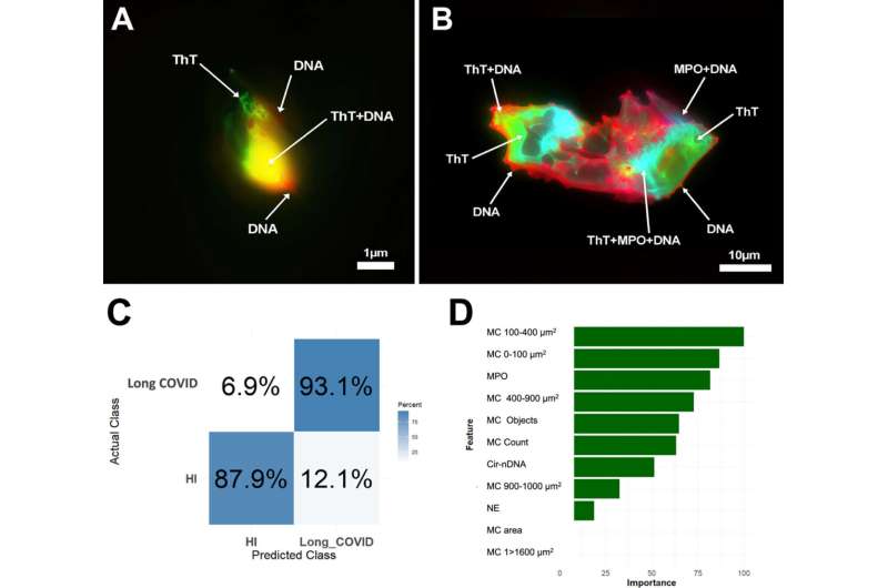
Illustrative co-localization of NETs and microclots markers in control and LC individual plasma, and performance of LC quantitative detection performance. Credit:Journal of Medical Virology(2025). DOI: 10.1002/jmv.70613
In patients with long COVID, anew studyhas revealed a structural association between circulating microclots and neutrophil extracellular traps (NETs).
This finding suggests the existence of underlying physiological interactions between microclots and NETS that, when dysregulated, may become pathogenic. The work is published in theJournal of Medical Virology.
The term microclots, recently adopted in the scientific literature, refers to abnormal clumps of blood clotting proteins circulating in a patient's bloodstream. The concept was introduced in2021by Prof Resia Pretorius from Stellenbosch University's Department of Physiological Sciences, when they found the abnormal presence of suchmicroclotsin the blood samples of COVID-19 patients. Thisdiscoverygenerated widespread attention during the pandemic due to its potential role in COVID-related coagulopathies.
Dr. Alain Thierry's team at the Montpellier Cancer Institute (IRCM) at INSERM in Montpellier, was among the first to identify the critical role of NETs in the pathogenesis of COVID-19. NETs are produced through a specialized form of innate immune response known as NETosis, whereby neutrophils expel their DNA to form filamentous structures embedded with cytotoxic enzymes capable of rapidly trapping and neutralizing pathogens.
However, excessive NETs formation can become detrimental, contributing to a wide range of inflammatory and thrombotic diseases, including severe infections, autoimmune disorders, cancer, diabetes, and arthritis.
According to Dr. Thierry, it may be that persistent overproduction of NETs, fueled by self-perpetuating inflammatory and thrombotic loops, exacerbates disease severity.
In a collaborative effort, the teams of Prof Pretorius and Dr. Thierry investigated the potential interaction between microclots and NETs in the context of long COVID.
Using imagingflow cytometryandfluorescence microscopy, they performed a quantitative and structural analysis of microclots and NETs in the plasma of long COVID patients, compared to healthy controls. NETs were also quantified by analyzing NETs proteic markers and circulating DNA.
"This finding suggests the existence of underlying physiological interactions between microclots and NETs that, when dysregulated, may become pathogenic," explains Dr. Thierry.
Furthermore, the integration of Artificial Intelligence tools, such asmachine learning, into the biomarker analysis enabled them to distinguish long COVID patients from healthy individuals with high accuracy. The algorithms identified the most predictive biomarker combinations, enhancing diagnostic reliability and paving the way for personalized medicine approaches.
According to Prof Pretorius, the results reveal a significant accumulation of microclots in the plasma of long COVID patients, likely driven and stabilized by excessive NETs production: "This interaction could render microclots more resistant to fibrinolysis, promoting their persistence in circulation and contributing to chronic microvascular complications," she explains.
By identifying the mechanistic role of NETs in microclot stabilization, this study provides new insight into the pathophysiology of long COVID. These findings support the development of targeted therapeutic strategies aimed at modulating thrombo-inflammatory responses.
Finally, the study paves the way for the development of novel biomarkers for diagnosis and management: "The combination of advanced imaging techniques and machine learning confers methodological robustness and contributes significantly to the ongoing scientific discourse on post-viral syndromes," they conclude.
More information: Alain R. Thierry et al, Circulating Microclots Are Structurally Associated With Neutrophil Extracellular Traps and Their Amounts Are Elevated in Long COVID Patients, Journal of Medical Virology (2025). DOI: 10.1002/jmv.70613
Provided by Stellenbosch University


Post comments