
byOptica
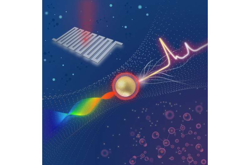
Researchers developed a sensitive Raman imaging system that can detect very faint signals from SERS nanoparticles that bind to tumor markers. Credit: Zhen Qiu, Michigan State University
Researchers have developed a new compact Raman imaging system that is sensitive enough to differentiate between tumor and normal tissue. The system offers a promising route to earlier cancer detection and to making molecular imaging more practical outside the lab.
The new Raman system is designed to detect very faint signals from specialsurface-enhanced Raman scattering(SERS) nanoparticles that bind to tumor markers. After the particles are applied to a sample or the area being examined, the imaging system reads their signal and automatically highlights spots that are likely to contain tumor tissue.
"Traditional methods for cancer-related diagnosis are time-consuming and labor-intensive because they require staining tissue samples and having a pathologist look for any abnormalities," said research team leader Zhen Qiu from the Institute for Quantitative Health Science and Engineering (IQ), Michigan State University.
"While our system would not immediately replace pathology, it could serve as a rapid screening tool to accelerate diagnosis."
InOptica, Qiu and colleagues describe their new imaging system and show that it can distinguish cancerous from healthy cells while detecting Raman signals about four times weaker than a comparable commercial system.
They achieved this sensitivity by combining aswept-source laser—which changes wavelength during analysis—with an ultra-sensitive detector known as a superconducting nanowire single-photon detector (SNSPD).
"This technology could eventually enable portable or intraoperative devices that enable clinicians to detect cancers at earlier stages, improve the accuracy of biopsy sampling and monitor disease progression through less invasive testing," said Qiu.
"Ultimately, such advances could enhance patient outcomes and reduce diagnostic delays, accelerating the path from detection to treatment."

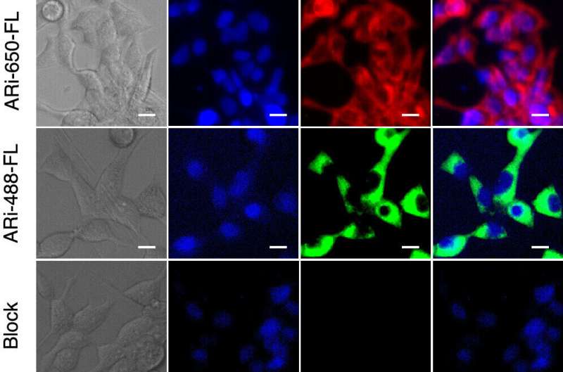
The researchers used the Raman system to image breast cancer cells incubated with SERS nanoparticles coated with hyaluronan acid, which causes the nanoparticles to bind to a surface protein expressed in many tumor cells. (a) Wield-field image of cancer cells, with a red dashed box representing the scanning region. (b) Overlap image of SERS intensity map and wide-field image of the breast cancer cells. Points A and B mark representative locations on a targeted cell and background region, respectively. (c) Representative Raman signal from the cell and background highlighted in (b). Scale bar: 20 microns. Credit: Zhen Qiu, Michigan State University
Qiu's lab explores how SNSPDs can be used to improve various imaging platforms. SNSPDs use a superconducting wire to detect individual particles of light, making it possible to capture extremely weak optical signals with high speed and very low noise.
For this project, they wanted to use an SNSPD to create a platform that detects Raman signals far weaker than those measurable with current Raman systems. Raman imaging maps the chemical makeup of a sample by measuring the unique light-scattering fingerprints of its molecules. These signals can be amplified by using SERS nanoparticles.
"Combining this advanced detector with aswept‑source Raman architecturethat replaces a bulky camera and collects light more efficiently resulted in a system with a detection limit well beyond that of comparable commercial systems," said Qiu.
"Also, the fiber coupling configuration and compact design facilitate system miniaturization and future clinical translation."
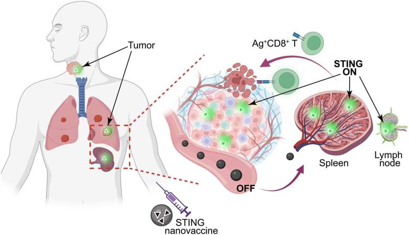
The researchers evaluated the new system usingSERS nanoparticlesthat were coated with hyaluronan acid, which causes the nanoparticles to bind to CD44, a surface protein expressed in many tumor cells.
They began by testing simple solutions of SERS nanoparticles, showing that the imaging system could achieve femtomolar sensitivity. They then used the system to examine cultured breast cancer cells, mouse tumors and healthy tissues.
"The SERS signals were strongly concentrated in tumor samples, with only minimal background detected in healthy tissue," said Qiu.
"This demonstrates both the system's exceptional sensitivity and its ability to provide reliable tumor‑versus‑healthy contrast. Moreover, by adjusting or substituting the targeting molecule, this method could be adapted for other cancer types."
According to the researchers, translating the system into one suitable for clinical use would require a faster readout and broader validation. They are now working to improve the imaging system's speed by using alternative laser sources, such as VCSELs, or by narrowing the sweep range. They also plan to conductmultiplexing experimentswith different nanoparticles that target multiple biomarkers simultaneously.
More information Yifan Liu et al, High-Sensitivity SNSPD-Enabled Swept Source Raman Spectroscopy for Cancer Detection with SERS Nanoparticles, Optica (2025). DOI: 10.1364/optica.569117 Journal information: Optica
Provided by Optica


Post comments