
by Luke Auburn,University of Rochester
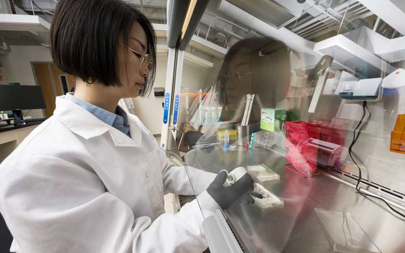
Chipping in: Biomedical engineering Ph.D. student Kaihua Chen, a member of URochester professor James McGrath's team, prepares tissue chips for experiments. Credit: University of Rochester photo / J. Adam Fenster
In lieu of animal experiments, researchers from the University of Rochester are using state-of-the-art microchips with human tissue to better understand how the brain operates under healthy conditions and is damaged through neurodegenerative diseases or conditions like sepsis.
James McGrath, the William R. Kenan Jr. Professor of Biomedical Engineering and director of the Translational Center for Barrier Microphysiological Systems (TraCe-bMPS), leads a team that develops and leverages tissue chips to study diseases where two different types of tissue meet, including at theblood-brain barrier. A pair of recent studies published inAdvanced ScienceandMaterials Today Bioused the chips to identify how the blood-brain barrier breaks down under serious threats, which could lead to new treatments to keep brains healthy.
When a patient undergoes a major surgery or contracts an infection such as sepsis, it can excessively inflame organs throughout the body including the brain, sometimes leading to long-lasting cognitive impairment, especially inolder patients.
In a study published inAdvanced Science, McGrath's team used tissue chips to show what happens at the barrier when the body suffers a cytokinetic storm—when theimmune systemcreates an uncontrollable systemic inflammatory response. Their experiments showed that with a high enough cytokine storm, the blood-brain barrier breaks down, leading tobrain injury.
"Two different stress signals—blood proteins that leak into the brain, like fibrinogen, together with inflammatory cytokines—can work together to trigger harmful changes in brain support cells called astrocytes," says Kaihua Chen, a biomedical engineering Ph.D. student and lead author of the study.
"At the same time, we found that the natural force of blood flow helps the blood-brain barrier stay stronger against these challenges. To me, this shows how both biology and engineering principles can come together to give us new insights into how the brain protects itself—and what goes wrong in disease."
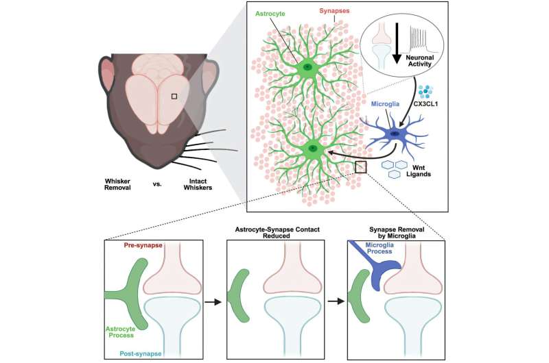
McGrath says that in the future, the team hopes to integrate more components of the brain on the brain side of the chip, including critical immune cells in the brain known as the microglia, to better understand how neurons are damaged during these inflammatory events. Ultimately, he hopes the chips can be used to prevent brain injuries in patients undergoing cytokine storms.
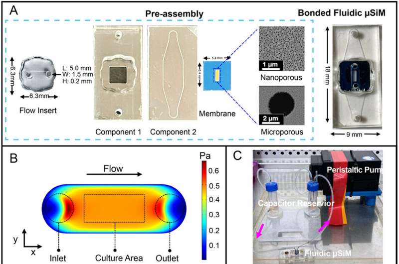
Development of fluidic µSiM-BBB. Credit:Advanced Science(2025). DOI: 10.1002/advs.202508271
"We hope that by building thesetissue modelsin chip format, we can arrange many brain models in a high-density array to screen candidates for neuroprotective drugs and develop brain models with diverse genetic backgrounds, including those that may be vulnerable or resilient to cytokine storms," says McGrath.
The researchers also envision their models being used in personalized medicine, tailored to individual patients' needs.
"If a patient is about to undergo a chemotherapy or amajor surgerythat risks generating cytokine storm, a chip modeling that specific patient's brain tissue could be used to evaluate risk and guide drug choice and dosing to help prevent brain injury as an outcome," McGrath says.
A second study, published inMaterials Today Bio, looked at pericytes, which are support cells that play an important but still not fully understood role in maintaining the blood-brain barrier. Previous studies have shown that in cases of systemic inflammation andneurodegenerative diseases, there are far fewer pericytes than in healthy brains, but it was not fully known why.
McGrath's team engineered holes and defects in endothelial tissue—the groups of cells that form blood vessels— and introduced pericytes to see what would happen.
"It's difficult forendothelial cellsto create a proper barrier when they're dealing with these large holes," says McGrath. "When we add the pericytes to the membrane, they create a beautiful matrix of structural fibers that fill those holes so the endothelial cells can make their vital barrier function."
Demonstrating the interaction between pericytes and endothelial cells opens the door to therapeutics that can preserve or introduce more pericytes to help keep the blood-brain barrier stable.
"By creating defects in the endothelial cell layer, we're letting the cells interact more directly, allowing the pericytes to provide some of the support they do in the body," says Michelle Trempel, a biomedical engineering Ph.D. student and lead author of the study. "This is important becausepericyteloss is implicated in many neurodegenerative diseases, so having a model where pericytes are providing support lets us study the impact of pericyte loss in the future."
More information: Kaihua Chen et al, Shear Conditioning Promotes Microvascular Endothelial Barrier Resilience in a Human BBB‐on‐a‐Chip Model of Systemic Inflammation Leading to Astrogliosis, Advanced Science (2025). DOI: 10.1002/advs.202508271 Michelle A. Trempel et al, Pericytes repair engineered defects in the basement membrane to restore barrier integrity in an in vitro model of the blood-brain barrier, Materials Today Bio (2025). DOI: 10.1016/j.mtbio.2025.102361 Journal information: Advanced Science
Provided by University of Rochester


Post comments